Diagnosing COVID-19 using Chest X-ray

COVID-SWIFT is a free Whatsapp based service to provide a swift diagnosis of potential COVID19 patients by analyzing Chest X-Ray images. Our state-of-the-art deep learning model generates a report containing predictions for COVID-19 and 14 other lung abnormalities with interpretable semantic markings on chest X-Ray. This can help doctors understand the severity of illness of their patients. We ran a small scale pilot for the last 10 months, where interested doctors within minutes, could receive a machine generated X-ray Report on sending us chest X-Ray of suspicious patients.


Our model is trained using Multi-Task Learning on multiple chest X-Ray datasets by NIH, RSNA, etc. We will soon be sharing our paper with further details.


COVID-SWIFT has now been launched as XraySetu in collaboration between IISc, Niramai, and ARTPARK. Xraysetu is quick and simple for busy doctors to use. XraySetu allows doctors in rural areas to plan early intervention for their patients by simply taking a picture of their Xray and sending it over via Whatsapp. We believe that this could be the model for the future of Indian healthcare, accessible to everyone wherever one might be.

Whatsapp based service

Send us Chest X-Ray at www.xraysetu.com to get patient report

Immediate Patient Report

Get instant patient report identifying presence of Pneumonia Type and Corona Virus

Locate COVID-19

Get regions of lungs infected with Pneumonia and Corona Virus

Covid Cases

And More Reports Generated

Sec for one patient report

Chest X-Ray used for Training


Patient Report Generation

Below images show sample report of patient tested COVID+ve
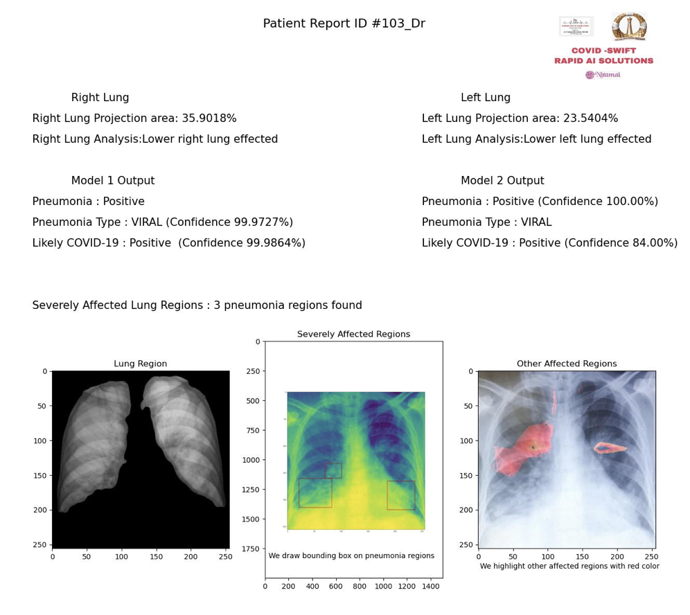
PATIENT REPORT

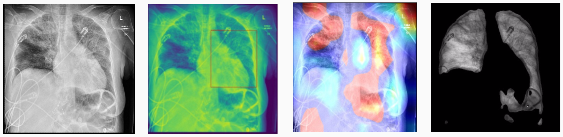
Chest X-Ray Analysis

The above image shows different stages of Chest X-Ray analysis our AI model interprests during identifying regions of high infections. (Left to Right) First image shows Chest X-Ray of COVID+ve patient. Second image consists of red bounding box which shows region of the lung where Pneumonia exists. Thind image gives suspecious regions of the lungs of the patient(red indicats regions of hight interest). Fourth image shows segmented lung region.

PROJECT INITIATIVE BY

Indian Institute of Science

NIRAMAI HEALTH Analytix

cardiology at doorstep